|
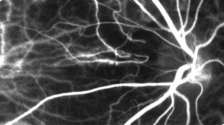
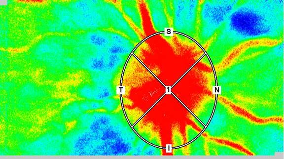
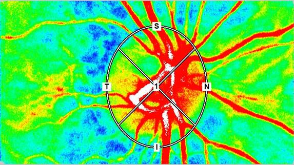
Branch Retinal Vein Occlusion(BRVO)
|
|
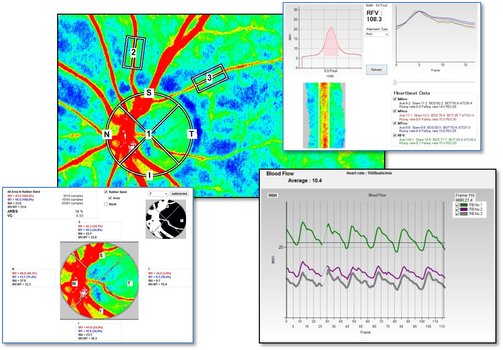
Composite Map
|
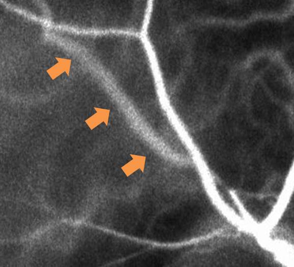
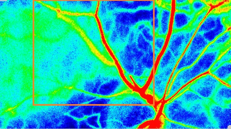
Enlarged Composite Map
(Gray Scale)
|
|
Images courtesy of Department of Ophthalmology, Tokyo Women's Medical University.
|
|
English papers related to BRVO
- NOMA, Hidetaka, et al.
Changes of retinal flow volume after intravitreal injection of bevacizumab in branch retinal vein occlusion with macular edema: a case series.
BMC ophthalmology, 2016, 16.1: 1.
- NITTA, Fumihiko, et al.
The effect of intravitreal bevacizumab on ocular blood flow in diabetic retinopathy and branch retinal vein occlusion as measured by laser speckle flowgraphy.
Clinical ophthalmology (Auckland, NZ), 2014, 8: 1119.
- MAENO, T., et al.
Measurement of Retinal Blood Flow Volume by Laser Speckle Flowgraphy Before and After Arteriovenous Sheathotomy for Branch Retinal Vein Occlusion.
Investigative Ophthalmology & Visual Science, 2008, 49.13: 4265-4265.
|
|
Central Retinal Vein Occlusion(CRVO)
|
|
Composite Map(before treatment)
|
Composite Map(after treatment)
|
|
Images courtesy of Department of Ophthalmology, Nagasaki University.
|
|
English papers related to CRVO
- NAGASATO, Daisuke, et al.
Correlation between optic nerve head circulation and visual function before and after anti-VEGF therapy for central retinal vein occlusion: prospective, interventional case series.
BMC ophthalmology, 2016, 16.1: 1.
- YAMADA, Yoshihisa, et al.
Retinal blood flow correlates to aqueous vascular endothelial growth factor in central retinal vein occlusion.
Retina, 2015, 35.10: 2037-2042.
- MATSUMOTO, Makiko, et al.
Retinal blood flow levels measured by laser speckle flowgraphy in patients who received intravitreal bevacizumab injection for macular edema secondary to central retinal vein occlusion.
Retinal Cases and Brief Reports, 2014, 8.1: 60-66.
- MAEDA, Kimihito; ISHIKAWA, Futoshi; OHGURO, Hiroshi.
Ocular blood flow levels and visual prognosis in a patient with nonischemic type central retinal vein occlusion.
Clinical ophthalmology (Auckland, NZ), 2009, 3: 489.
- TANO, R., et al.
Analysis of Retinal Blood Flow by Laser Speckle Flowgraphy in a Case With Central Retinal Vein Occlusion After Radial Optic Neurotomy.
Investigative Ophthalmology & Visual Science, 2008, 49.13: 4274-4274.
|