")
")
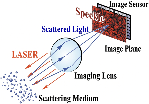
As shown in the figure, when living tissue is illuminated with a laser, scattered light randomly interferes with each other, generating speckles throughout the area. As shown in the figure, when the living tissue is imaged onto a screen through a lens, fine speckles also appear within this image. The speckle pattern at each point on this image plane changes moment by moment in accordance with the movement of blood cells near the corresponding point on the object plane—that is, the living tissue. By placing an image sensor on this image plane and calculating the rate of temporal change of the speckle pattern for each point, and displaying this as a map, it becomes possible to visualize blood flow in living tissue.