")
")
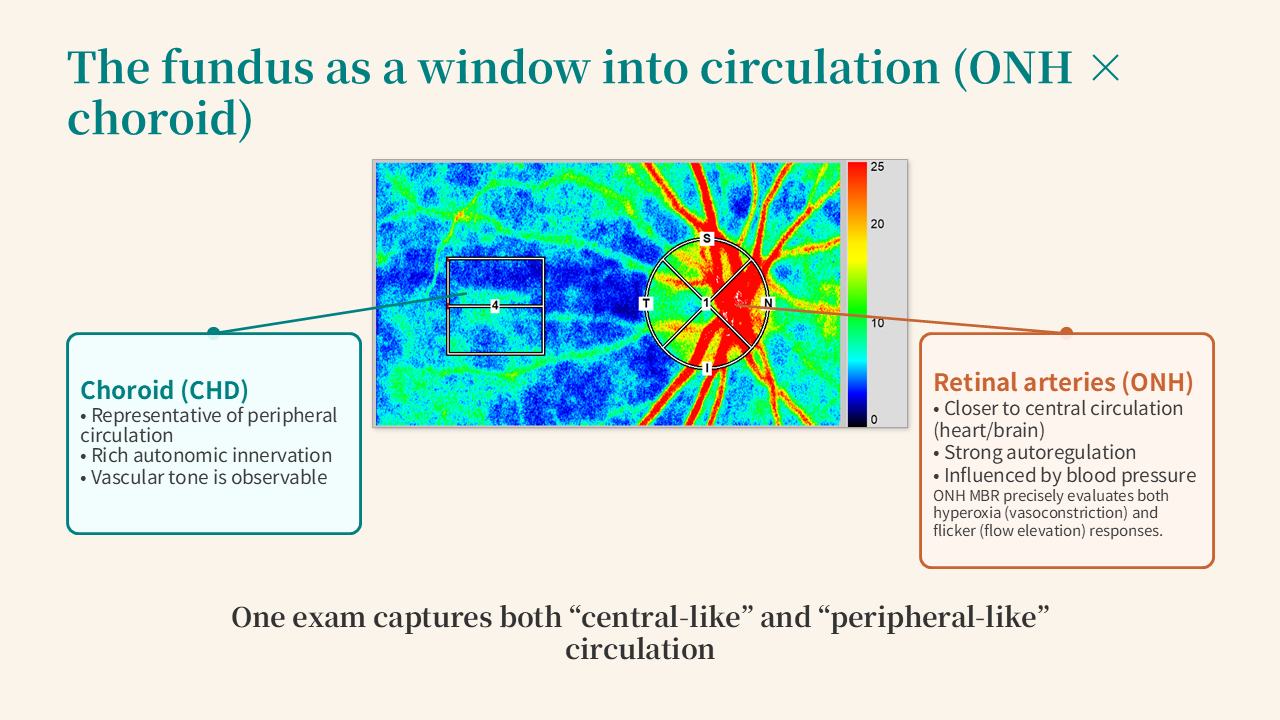

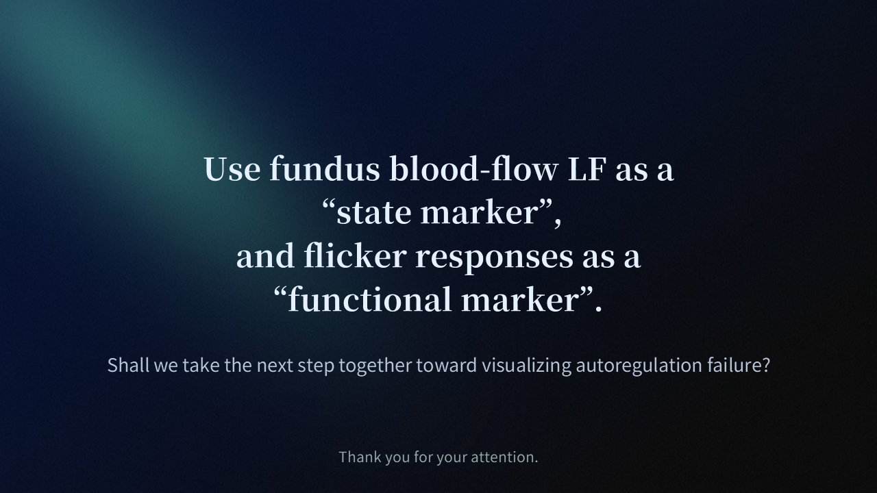
Explore a breakthrough in microvascular assessment with "Reading Autoregulation Through Retinal Blood Flow." Designed for researchers and CROs, this presentation reveals how the fundus serves as a non-invasive window into systemic circulation. Using Laser Speckle Flowgraphy (LSFG), we introduce a novel 4-phase model to evaluate neurovascular coupling and functional flow elevation. By focusing on the post-stimulation recovery trajectory, this methodology offers precise, quantifiable biomarkers for autoregulatory dysfunction and ischemia, empowering your clinical trials and neurovascular research.
Message for you,
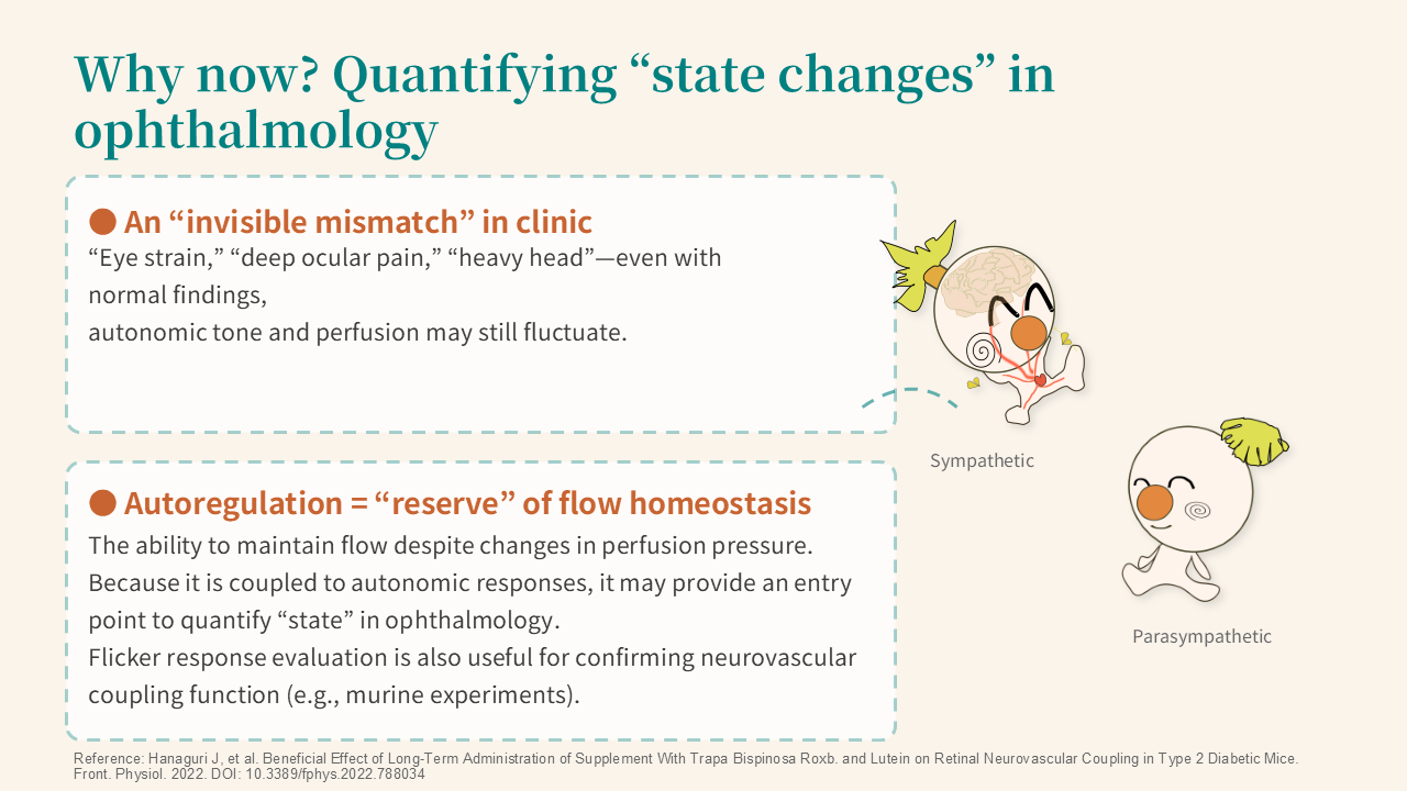
Many patients report eye strain, deep ocular pain, or a heavy sensation in the head — yet standard examinations reveal no abnormalities. This "invisible mismatch" may reflect fluctuations in autonomic tone and ocular perfusion that conventional static measurements cannot capture.
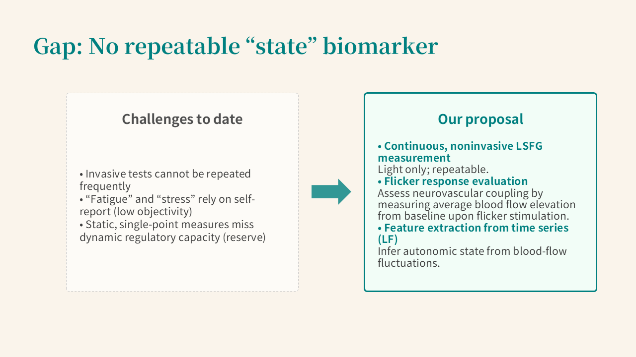
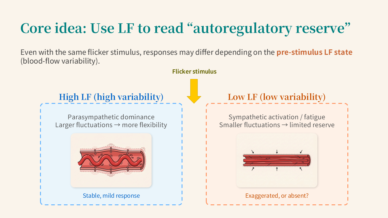
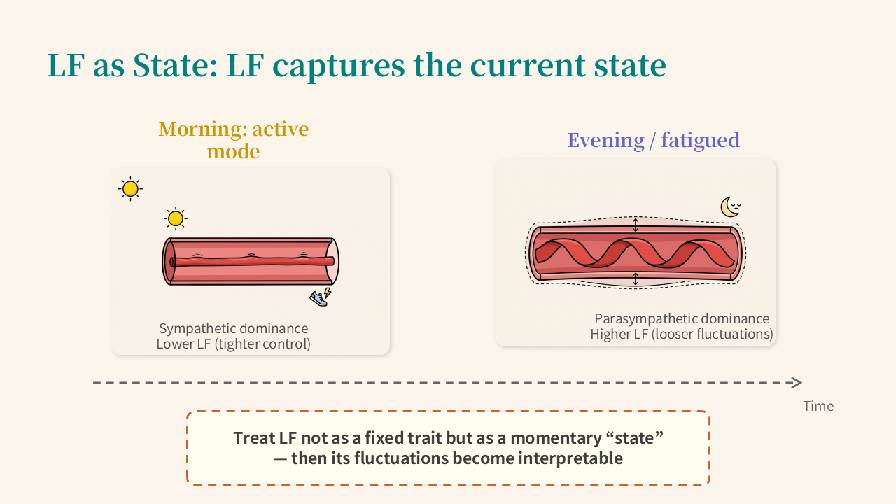
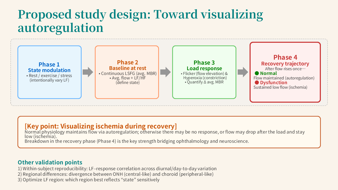
This presentation proposes using the low-frequency (LF) component of retinal blood flow, measured continuously via Laser Speckle Flowgraphy (LSFG), as a real-time autonomic state marker. LF shifts with time of day, fatigue, and stress — lower under sympathetic dominance, higher under parasympathetic dominance — and is interpreted here as a momentary "state" rather than a fixed trait.
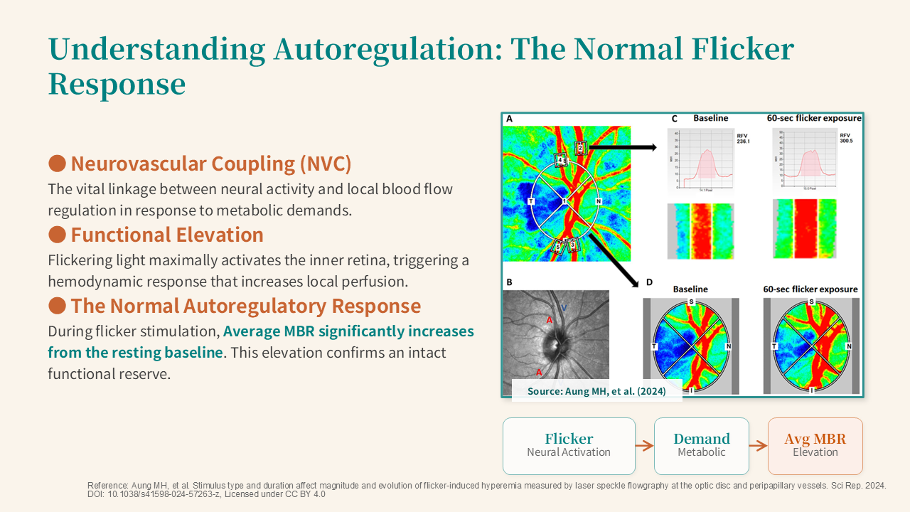
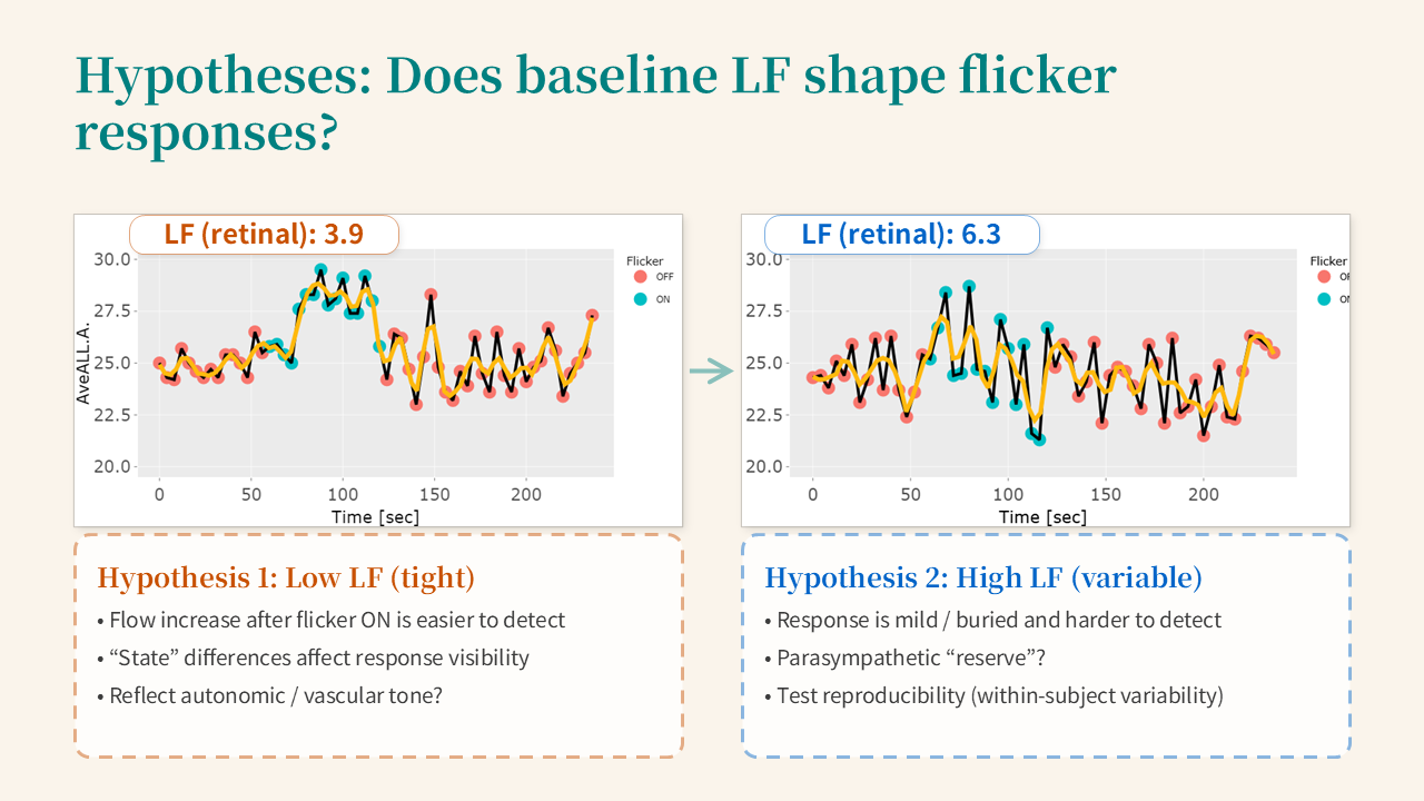
The central hypothesis is that the same flicker stimulus produces different blood flow responses depending on the pre-stimulus LF state. The recovery phase after stimulation is particularly telling: healthy autoregulation maintains elevated flow, while dysfunction may cause a sustained drop, potentially indicating ischemia.
If validated, this LF × flicker protocol could serve as a quantitative biomarker extending beyond ophthalmology into neurology, neuroscience, and systemic medicine.