")
")
In a word, it is a numerical value representing the resistance of retinal blood flow within the eye. A higher TCR indicates higher retinal blood flow resistance.
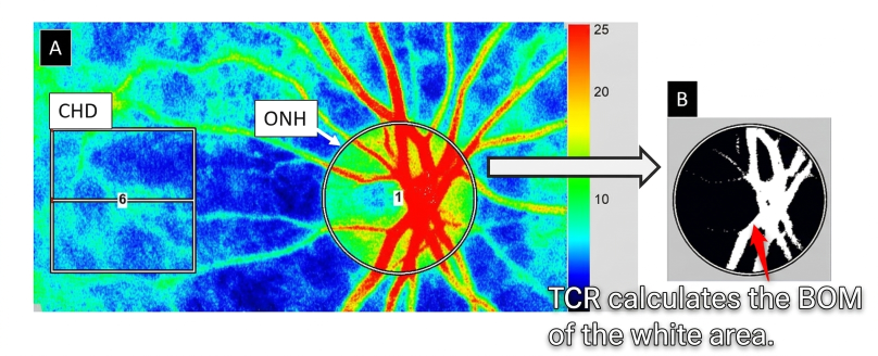
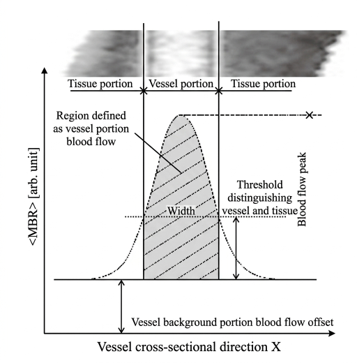
In LSFG, BOM exists as a new resistance index that does not require heartbeat detection. BOM quantifies the resistance of blood flow at any given location, and TCR (Total Capillary Resistance) is the BOM of the large vessel area (including arteries and veins) on the optic nerve head.
In this way, TCR represents the difference in resistance between blood flow measured from the large vessels of the optic nerve head (ONH) and the capillaries, and is a parameter used to estimate retinal vascular resistance.
Clinical applications include CRVO2 and glaucoma3, among others.
- Application in CRVO
The graph below is an excerpt from a published paper2 on CRVO cases. It summarizes the differences in TCR among healthy subjects, the affected eyes of CRVO patients, and the fellow eyes.
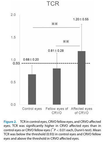
It can be seen that TCR in the affected eyes of CRVO is significantly higher compared to healthy eyes and fellow eyes. Furthermore, a cutoff value for TCR between healthy subjects and CRVO eyes (= 0.93) has been reported, suggesting that TCR can serve as a good indicator for CRVO.
- Application in Glaucoma
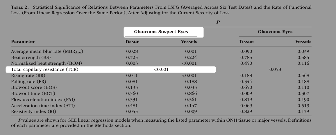
According to the literature3, when TCR is high, retinal vessels tend to be stiffer, and this stiffness has been suggested to be associated with progressive visual function decline. In particular, TCR is considered an important factor indicating the risk of progressive visual function decline. However, high TCR is not always strongly associated with the degree of existing visual function decline, and this is considered useful for risk assessment at an early stage.
Recent Reports
- Kenji Okamoto, Noriyoshi Takahashi, Tatsuhiko Kobayashi, Tomoaki Shiba, Yuichi Hori and Hitoshi Fujii
Novel superpixel method to visualize fundus blood flow resistivity in healthy adults
Scientific Reports, 13, doi:10.1038/s41598-023-33450-2, 2023. - Makiko Matsumoto, Kiyoshi Suzuma, Fumito Akiyama, Kanako Yamada, Shiori Harada, Eiko Tsuiki and Takashi Kitaoka
Retinal Microvascular Resistance Estimated from Waveform Analysis Is Significantly Higher With a Threshold Value in Central Retinal Vein Occlusion
Translational Vision Science and Technology, 9(11), 4, doi:10.1167/tvst.9.11.4, 2020. - Stuart Keith Gardiner, Cindy Albert, Brad Fortune, Steven L Mansberger and Grant Cull
Retinal vessel pulsatile characteristics can predict functional progression in glaucoma suspects
Investigative Ophthalmology and Visual Science, 64(8), 3778, 2023 ARVO Annual Meeting abstract, 2023.